In a new paper published this month in Applied Animal Behavior Science, recent MS graduate Kira Lemke demonstrates that unlike octopuses, cuttlefish don’t engage in play with objects.
Welfare standards for cephalopods are generally poorly supported by rigorous studies. Enclosure enrichments are variable, and whether enrichments actually meet needs of different cephalopod taxa is not widely studied. “Enrichment” also has multiple meanings and different applications, including anything from substrate on the enclosure floor, provision of live prey, puzzle boxes, novel objects, housing rearrangement, human interaction and many others. And while there have been a number of studies demonstrating that octopuses in research laboratories and public aquaria engage in extended play-like behavior with novel objects, it is unknown whether these provide a genuine welfare benefit, or whether other cephalopods also display similar behaviors.
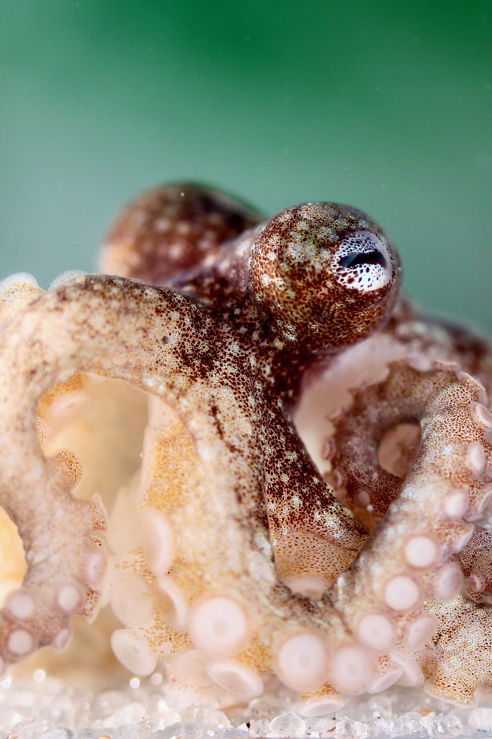
In this study based on her thesis work, Kira demonstrates conclusively that dwarf cuttlefish (Sepia bandensis) do not interact with novel objects placed into their housing enclosures, and that this lack of engagement is consistent whether their housing environment contains static enrichment or not. This paper indicates that species- and taxon-specific enrichments need to be assessed and implemented based on the needs of each different cephalopod clade, and also that high levels of cognitive ability (which are certainly present in cuttlefish) do not necessarily correlate with play-like behaviors.
Congratulations to Kira on the publication of an important and timely study!